RIKEN Center for Life Science Technologies

Topics

  • Research

    Latest publications from CLST.
     
  • News

    Awards, agreements, and other news.
  • Events

    Upcoming and past events including seminars, symposiums, open days.

For Public

To let you know about our research, this area contains 4 types of information about CLST; “Article”, “Videos”, “Event” and “Study”.
At “Article”, you can read articles on interviews and lectures, and you can enjoy the videos about CLST at “Videos”. If you want to meet and talk directly with the researcher, “Visit” give you some information of such events. You can find more difficult contents to know about our research deeply at “Study”.
  • Article
  • Event
  • Videos
  • Study

Labs & Technologies

Cellular Dynamics Analysis Unit

Biomolecular functions highlighted by leading-edge light microscopy technologies


* Due to the reorganization starting as new centers in April 2018, this laboratory is now belong to the Center for Biosystems Dynamics Research. As for the latest information, please see the following URL below.
> The webpage of Laboratory for Molecular and Cellular Dynamics, Center for Biosystems Dynamics Research

Unit Leader
Yuko Mimori-Kiyosue  Ph.D.

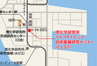
2-2-3 Minatojima-minamimachi, Chuo-ku, Kobe, Hyogo 650-0047, Japan
Tel: +81-78-306-3224

3_14_kiyosue.png

>>> Lab Site

Research Area

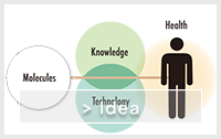
We aim to elucidate the functions of biomolecules supporting vital activities and thereby contribute to extend the human health span. Activities of every single cell that forms the living body are supported by intracellular infrastructure. We particularly focus on the molecular mechanisms that control the cellular infrastructure “microtubules” responsible for intracellular trafficking, and analyze their function using cell models and mouse models, while making full use of the common-use Light Microscopy Facility that our laboratory run.


1) Functional analysis of intracellular machinery driving the life

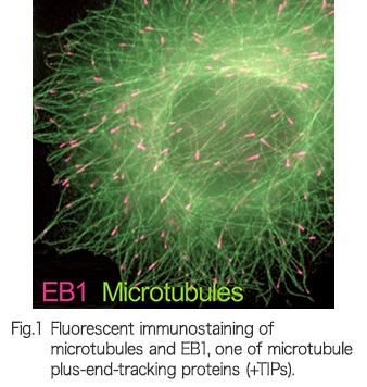
Appropriate organization of the microtubule cytoskeleton is required for intracellular transport and chromosome segregation. Its fine-tuning is critical for normal cell motility, morphogenesis and cell division, while the abnormal control is the cause of the disease [2-10]. We study how the activities of microtubule regulating molecules (Fig.1) support normal development or disease through phenotypic analysis of mouse and in vitro cell biology, to better understand the mechanisms of life and at the same time contribute to medical science.


2) Construction of Lattice Light Sheet Microscope and 3D analysis of cellular dynamics

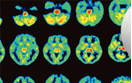
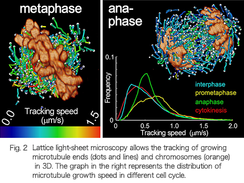
The Lattice Light Sheet Microscope developed by Dr. Betzig laboratory (Howard Hughes Medical Institute) is a breakthrough technology overcoming the limitation of conventional fluorescent microscopy, which achieves whole cell 3D scan within one second at high spatiotemporal resolution [1]. Using this system, we have succeeded in capturing 3D dynamics of microtubule growth and chromosome movement in unprecedented accuracy (Fig.2). We are advancing efforts toward constructing this microscope in our lab to realize highly accurate 3D analysis of cellular events including cell division.


3) The management of institute's common-use imaging facility

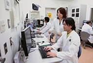
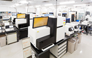
 We run common-use Riken Kobe LIGHT MICROSCOPY FACILITY (kLMF), which offers about twenty light microscope system products and image analysis software.

Main Publications List

1

Three-dimensional tracking of plus-tips by lattice light-sheet microscopy permits the quantification of microtubule growth trajectories within the mitotic apparatus

Yamashita N, Morita M, Legant WR, Chen BC, Betzig E, Yokota H, Mimori-Kiyosue Y
J Biomed Opt., 20(10), 101206 (2015).
2

Lattice light-sheet microscopy: imaging molecules to embryos at high spatiotemporal resolution.

Chen BC, Legant WR, Wang K, Shao L, Milkie DE, Davidson MW, Janetopoulos C, Wu XS, Hammer JA rd, Liu Z, English BP, Mimori-Kiyosue Y, Romero DP, Ritter AT, Lippincott-Schwartz J, Fritz-Laylin L, Mullins RD, Mitchell DM, Bembenek JN, Reymann AC, Böhme R, Grill SW, Wang JT, Seydoux G, Tulu US, Kiehart DP, Betzig E
Science, 346(6208), 1257998 (2014).
3

Improving spinning disk confocal microscopy by preventing pinhole cross-talk for intravital imaging.

Shimozawa T, Yamagata K, Kondo T, Hayashi S, Shitamukai A, Konno D, Matsuzaki F, Takayama J, Onami S, Nakayama H, Kosugi Y, Watanabe TM, Fujita K, Mimori-Kiyosue Y.
Proc Natl Acad Sci U S A, 110(9), 3399-3404 (2013).
4

Dissecting the nanoscale distributions and functions of microtubule-end-binding proteins EB1 and ch-TOG in interphase HeLa cells.

Nakamura S, Grigoriev I, Nogi T, Hamaji T, Cassimeris L, Mimori-Kiyosue Y.
PLoS One, 7(12), e51442 (2012).
5

Shaping microtubules into diverse patterns: molecular connections for setting up both ends.

Mimori-Kiyosue Y.
Cytoskeleton (Hoboken), 68(11), 603-618 (2011).
6

Laminin-based cell adhesion anchors microtubule plus ends to the epithelial cell basal cortex through LL5α/β

Hotta A, Kawakatsu T, Nakatani T, Sato T, Matsui C, Sukezane T, Akagi T, Hamaji T, Grigoriev I, Akhmanova A, Takai Y, Mimori-Kiyosue Y
J Cell Biol, 189(5), 901-917 (2010).
7

CLASP1 and CLASP2 bind to EB1 and regulate microtubule plus-end dynamics at the cell cortex.

Mimori-Kiyosue Y, Grigoriev I, Lansbergen G, Sasaki H, Matsui C, Severin F, Galjart N, Grosveld F, Vorobjev I, Tsukita S, Akhmanova A.
J Cell Biol, 168(1), 141-153 (2005).
8

Search-and-capture of microtubules through plus-end-binding proteins (+TIPs).

Mimori-Kiyosue Y, Tsukita S.
J Biochem, 134(3), 321-326 (2003).
9

The dynamic behavior of the APC-binding protein EB1 on the distal ends of microtubules.

Mimori-Kiyosue Y, Shiina N, Tsukita S.
Curr Biol, 10(14), 865-868 (2000).

>>>ALL Publications

Member  *concurrent

CLST was reorganized into three centers according to the RIKEN 4th Medium-Term Plan from April 1, 2018. For the latest information of Cellular Dynamics Analysis Unit, please visit the following websites.


> The webpage of Laboratory for Molecular and Cellular Dynamics, Center for Biosystems Dynamics Research [http://www.bdr.riken.jp/en/research/labs/kiyosue-y/index.html]